


In the case of thoracic osteochondrosis, organs associated with areas of the spinal cord, which is located at the level of the affected thoracic region and below, often suffer. Violation of the normal activity of the spine leads to immobility of the arms, legs and torso as a whole, to dysfunction of the pelvic organs, respiratory muscles and internal organs.
Osteochondrosis is a degenerative-dystrophic disease of the spine, which is based on a change in the intervertebral discs with involvement in the pathological process of neighboring vertebrae and intervertebral joints with the entire ligament apparatus.
Features of the anatomy of the spine
The mobility and stability, elasticity and elasticity of the spine largely depend on the intervertebral discs, which are one of the types of cartilage connection between bones and provide a strong bond between the bodies of neighboring vertebrae. The total length of the intervertebral discs is one quarter of the length of the spinal column.
The most important function of the discs is to reduce the vertical load on the vertebrae. The disc consists of three parts:
- hyaline plates (closely adjacent to the vertebrae);
- nucleus pulposus (fills the space between the plates);
- fibrous ring (surrounds the nucleus from the outside).
The core contains cartilage cells, tightly woven collagen fibers and chondrin (proteoglycans). The anterior surface of the discs is covered by the anterior longitudinal ligament, which is tightly fused with the vertebrae and freely turns the discs over. The posterior longitudinal ligament is firmly fused with the disc surface and forms the anterior wall of the spinal canal. The intervertebral disc does not have its own blood supply, so it feeds on substances that come by diffusion from the vertebral bodies.
The distribution of vertical loads in the spine occurs due to the elastic properties of the discs. As a result of the pressure, the nucleus pulposus expands and the pressure is redistributed to the fibrous ring and hyaline plates. During movement, the core moves in the opposite direction: when flexed - towards convexity, when it is inflexible - anteriorly. When the spine moves, muscles, ligaments and discs are included in the work. Thus, a violation in one link leads to a violation of the entire kinetic chain.
Causes and mechanism of the development of the disease
In the development of osteochondrosis, a special role is played by the mechanical effect on the spine. Under the influence of unfavorable static and dynamic loads, the pulpy nucleus gradually loses its elastic properties (due to the depolymerization of polysaccharides), forms protrusions and sequesters.
The process of disc degeneration is influenced by a genetic predisposition, which provokes the development of changes in the neuromuscular apparatus of the back, a change in the structure of glycosamines and a violation of the distribution of collagen fibers in the disc. The genetic factor is fundamental in the onset of thoracic osteochondrosis, subject to greater functional activity.
Risk factors for developing degenerative changes in the spine include anatomical features of the discs, which are imperfections in evolution. One of these characteristics are the nutritional characteristics of the structures. In the human body, the disc is made up of poorly perfused tissue. The closure of blood vessels already occurs in childhood. After nutrition occurs due to the diffusion of substances through the end plates.
The stimulator of the penetration of nutrients is a dosed load that excludes static postures and great stress. Physical inactivity is a major risk factor for thoracic osteochondrosis. Therefore, regular exercise is an important preventive measure.
The peculiarity of the microscopic structure - few cells - reduces the intensity of the regenerative capacity and the recovery speed of the disc components. An anatomical feature is the weakness and lack of strength of the discs in the posterior sections. This contributes to the appearance of wedge-shaped discs in the lower thoracic and lumbar regions.
Great importance in the development of osteochondrosis is given to involutionary changes. Actively degenerative changes begin to increase after 30 years. The synthesis of the components necessary for the disc (glycosaminoglycans) continues, but their quality is deteriorating. Hydrophilicity decreases, fibrosity increases, sclerosis appears.
Stages of degeneration of the intervertebral discs:
- prolonged asymptomatic course, degenerative changes of the intradiscal components, displacement of the nucleus within the disc;
- pronounced root symptoms of thoracic osteochondrosis, compression of the spinal cord, protrusion of the nucleus pulposus (protrusion, 1 degree);
- rupture of the disc with hernial protrusion (hernia, 2nd degree);
- degenerative changes of the extradiscal components (grade 3).
Pathological protrusion compresses the nerve roots, blood vessels or the spinal cord at various levels (cervical, thoracic, lumbar), determining the clinical picture.
Restriction of mobility in the thoracic spine, due to the presence of the thorax, contributes to minimal traumatization of the intervertebral discs and therefore osteochondrosis. Physiological thoracic kyphosis contributes to the redistribution of the weight of the upper half of the body to the lateral and anterior sections of the vertebrae. Thus, intervertebral hernias and osteophytes form on the anterior and lateral surfaces of the spine. Posterior osteophytes and hernias are extremely rare.
Osteochondrosis contributes to the narrowing of the intervertebral foramina and compression of the spinal cord roots and sympathetic fibers. Sympathetic fibers originate in the gray matter of the spinal cord, then collect in nodes, from which they are sent to all internal organs. This leads to the fact that thoracic osteochondrosis, in addition to typical neurological disorders, leads to dysfunctions of the internal organs (vegetative, vasomotor, trophic) and the imitation of somatic diseases. This feature of osteochondrosis of the thoracic discs explains the difficulties in diagnosing and prescribing the correct treatment.
Thoracic osteochondrosis symptoms
Thoracic osteochondrosis is more typical for people with a sedentary lifestyle. At the same time, there is no stimulating effect of dosed loads on the spine, which contributes to the disruption of disc restoration. Diseases develop in people who work at the computer for a long time, stoop, etc. these people need to independently perform therapeutic exercises.
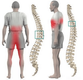
Most often, thoracic osteochondrosis is manifested by dull, less often aching and burning pains. The pain is localized between the shoulder blades. The patient is disturbed by the sensation of chest compression. When feeling the spinous processes of the thoracic vertebrae, local pain is detected, which increases with axial loads on the spine, deep inspiration and bends of the body.
A number of patients have severe pain in the shoulder blade and lower chest (posterior rib syndrome). This symptomatology develops due to the displacement of the lower ribs. The pain sharply increases when turning the torso. More often than not, the pain syndrome suddenly disappears.
Often the pain in the chest becomes girdle, corresponds to the course of the intercostal nerve. Sensitivity in the innervation zone of the corresponding nerve ending is disturbed, paresthesias appear, and often there is a decrease in superficial and deep sensitivity. Possible violation of the function of the abdominal press, a change in the reflexes of the knee and heel tendon.
Violation of the function of internal organs occurs when any nerve root is compressed at level 1 to 12 of the chest. In the thoracic region there are structures responsible for innervation of the lungs, heart, intestine, liver, pancreas and kidneys. Therefore, there are no characteristic signs only for thoracic osteochondrosis.
The disease manifests itself with the characteristic symptoms of another pathology:
- difficulty breathing;
- intense night pains;
- "heart", anginal pains;
- pain in the mammary glands;
- pain in the right or left hypochondrium (symptoms of cholecystitis and pancreatitis);
- pain in the throat and esophagus;
- pain in the epigastrium, abdomen (symptoms of gastritis, enteritis and colitis);
- sexual dysfunction.
Diagnostics
The greatest value in diagnosing thoracic osteochondrosis is a chest X-ray examination. The picture shows a decrease in the height of the intervertebral disc, sclerosis of the end plates, the formation of osteophytes.
Computed tomography allows you to clarify the condition of the vertebrae, joints of the spine, the size of the spinal canal, determine the location of the hernial protrusion and its size.
When performing differential diagnosis, it is necessary to carefully collect an anamnesis and compare all clinical signs of thoracic osteochondrosis with the symptoms of other diseases. For example: heart pain with osteochondrosis is not stopped by nitroglycerin, epigastric pain is not associated with food intake, is not seasonal, all symptoms appear mainly in the evening and completely disappear after a night's rest.
How to treat thoracic osteochondrosis?
Treatment of osteochondrosis of the thoracic spine in almost all cases is conservative. The indication for therapy is the predominance of visceral syndromes with neurological disorders. The main orthopedic treatment should be adequate traction of the spine:
- active vertical traction underwater;
- passive horizontal traction in an inclined bed using the Glisson loop in case of damage at the level of 1-4 thoracic vertebrae, from the axillary straps in case of damage at the level of 4-12 thoracic vertebrae.
Drug treatment consists in performing paravertebral blocks with a solution of novocaine. With an exacerbation of the disease, analgesics and sedatives are used. With an unexpressed pain syndrome, it is permissible to use ointments with analgesics and anti-inflammatory drugs at home.
After the elimination of acute phenomena, a massage of the muscles of the back and lower limbs is used. Manual therapy is indicated for 1-3 degrees of osteochondrosis in case of the development of functional blocks. Includes various options for soft and rough effects on the back muscles.
Therapeutic exercise allows you to load all parts of the spine in a measured way, stimulating the recovery processes. An important condition for physical therapy for osteochondrosis is to exclude vertical loads.
Physiotherapy: UHF treatments, ultrasounds, inductothermy, baths with radon salts and pine-conifers. In the thermal phase, underwater traction and hydromassage are actively used.
Surgical treatment is rarely used. The indication for surgery is compression of the spinal cord by a prolapsed disc fragment.